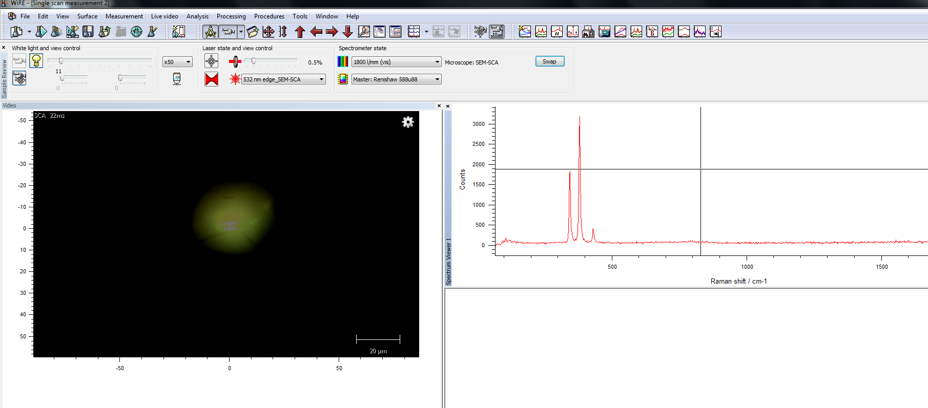
Combining Raman Microscopy With Scanning Electron Microscopy Sem To Raman imaging and scanning electron microscopy (rise) combine the advantage of scanning electron microscope and raman spectroscopy, which can collect the morphology, composition, and structure information in the same micro region of the geological sample in situ. Confocal raman imaging and scanning electron (rise) microscopy, when combined in a microscope, complement each other and provide the emerging opportunities to clarify morphological, structural, and chemical information of materials at the micron and even nanoscale.
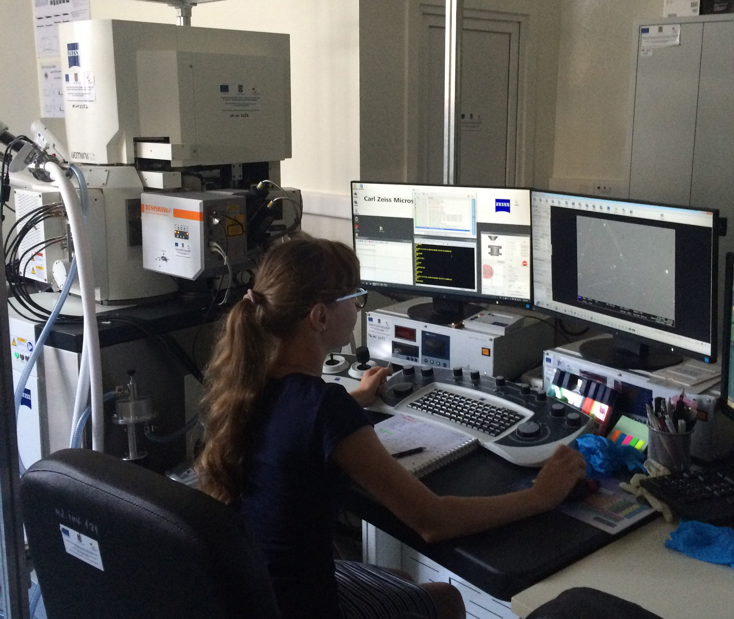
Combining Raman Microscopy With Scanning Electron Microscopy Sem To Advanced mapping by micro raman spectroscopy and scanning electron microscopy equipped with energy dispersive spectroscopy have been combined to disclose molecular and elemental features on the same regions sample at a micrometric resolution. Due to the specific vacuum requirements for scanning electron microscopy (sem), the raman microscope has to operate in vacuum in a correlative raman sem, which is a type of microscope combination that has recently increased in popularity. Rise microscopy is the combination of confocal raman imaging and scanning elec tron microscopy. it incorporates the sensitivity of the non destructive, spectroscopic raman technique along with the atomic resolution of electron microscopy. Raman imaging and scanning electron (rise®) microscopy combined with energy dispersive x ray spectroscopy (eds) offers comprehensive sample characterisation at the nanoscale. request pricing. rise instruments seamlessly integrate confocal raman imaging and scanning electron microscopy (sem).
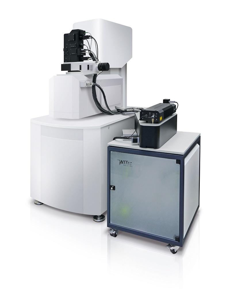
Correlative Raman Imaging And Scanning Electron Microscopy Raman Sem Rise microscopy is the combination of confocal raman imaging and scanning elec tron microscopy. it incorporates the sensitivity of the non destructive, spectroscopic raman technique along with the atomic resolution of electron microscopy. Raman imaging and scanning electron (rise®) microscopy combined with energy dispersive x ray spectroscopy (eds) offers comprehensive sample characterisation at the nanoscale. request pricing. rise instruments seamlessly integrate confocal raman imaging and scanning electron microscopy (sem). They use renishaw’s structural and chemical analyser (sca) interface to bring the raman analysis capabilities of the invia™ confocal raman microscope to their scanning electron microscope (sem). Correlative raman sem imaging (rise microscopy, witec, ulm, germany and tescan orsay holding, brno, czech republic) combines an sem and a confocal raman microscope. the confocal raman microscope is integrated into the vacuum chamber of the electron microscope. non destructive raman and sem measurements are consecutively performed at. Scanning transmission electron microscopy (stem) employs a specific type of detector installed on the sem to observe thin sections of materials in parallel with transmission electron microscopy (tem). sem can be combined with chemical microanalysis (energy dispersive x ray spectrometry eds, or wavelength dispersive x ray spectrometry wds). Although there is a wide range of analytical techniques to characterize polymeric materials, here the focus is laid on a novel correlative microscopy method, combining raman microscopy, scanning electron microscopy (sem) and energy dispersive x ray spectroscopy (edxs).
Correlative Raman Imaging And Scanning Electron Microscopy Raman Sem They use renishaw’s structural and chemical analyser (sca) interface to bring the raman analysis capabilities of the invia™ confocal raman microscope to their scanning electron microscope (sem). Correlative raman sem imaging (rise microscopy, witec, ulm, germany and tescan orsay holding, brno, czech republic) combines an sem and a confocal raman microscope. the confocal raman microscope is integrated into the vacuum chamber of the electron microscope. non destructive raman and sem measurements are consecutively performed at. Scanning transmission electron microscopy (stem) employs a specific type of detector installed on the sem to observe thin sections of materials in parallel with transmission electron microscopy (tem). sem can be combined with chemical microanalysis (energy dispersive x ray spectrometry eds, or wavelength dispersive x ray spectrometry wds). Although there is a wide range of analytical techniques to characterize polymeric materials, here the focus is laid on a novel correlative microscopy method, combining raman microscopy, scanning electron microscopy (sem) and energy dispersive x ray spectroscopy (edxs).
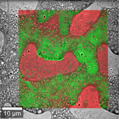
Correlative Raman Imaging And Scanning Electron Microscopy Raman Sem Scanning transmission electron microscopy (stem) employs a specific type of detector installed on the sem to observe thin sections of materials in parallel with transmission electron microscopy (tem). sem can be combined with chemical microanalysis (energy dispersive x ray spectrometry eds, or wavelength dispersive x ray spectrometry wds). Although there is a wide range of analytical techniques to characterize polymeric materials, here the focus is laid on a novel correlative microscopy method, combining raman microscopy, scanning electron microscopy (sem) and energy dispersive x ray spectroscopy (edxs).

Plan View Scanning Electron Microscopy Sem Micrographs And Raman
