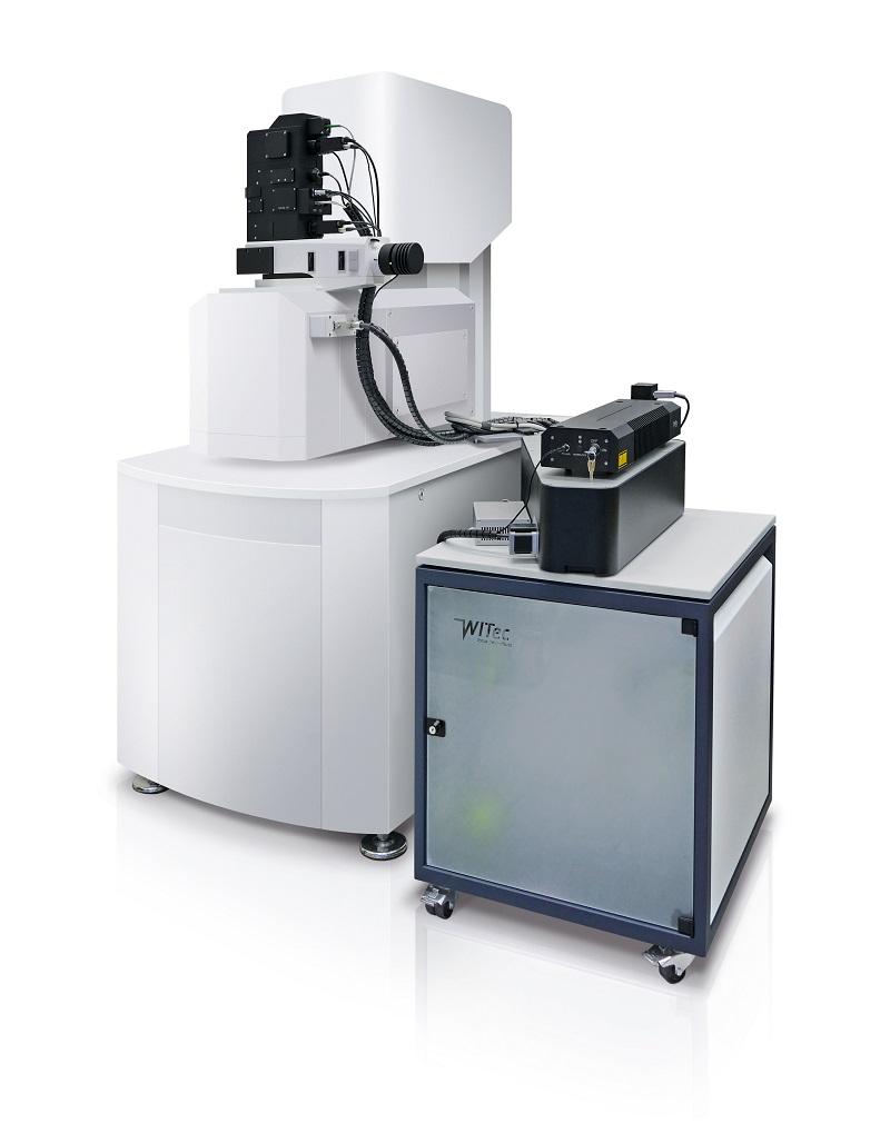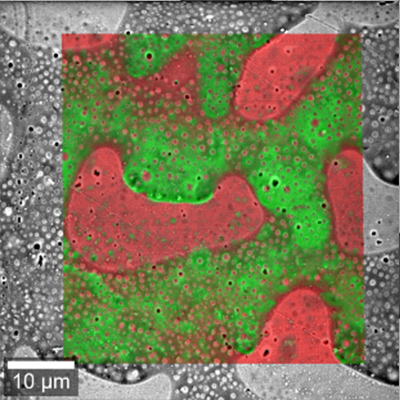
Correlative Raman Imaging And Scanning Electron Microscopy Raman Sem In this minireview, we summarize the principle of raman and the development of a correlative sem raman system and showcase the typical application of such rise microscopy in different fields. on this basis, the advantages of the rise system, comprising two complementary techniques, are highlighted. Correlative raman sem imaging (rise microscopy, witec, ulm, germany and tescan orsay holding, brno, czech republic) combines an sem and a confocal raman microscope. the confocal raman microscope is integrated into the vacuum chamber of the electron microscope. non destructive raman and sem measurements are consecutively performed at.

Correlative Raman Imaging And Scanning Electron Microscopy Raman Sem Fortunately, scanning electron microscopy (sem) can well compensate for the small depth of field and low spatial resolution of raman microscopy, achieving high resolution surface morphological images. sem has two basic and commonly used signals, secondary electron (se) and back scattered electron (bse). Here, we illustrate a workflow to correlate fish, sem, raman, and, if desired, nanosims to provide a comprehensive characterization of microorganisms at single cell resolution. Correlative raman imaging and scanning electron (rise) microscopy is a unique combination of scanning electron microscopy (sem) and confocal raman imaging. this innovative correlative microscopy technique enables linking ultra structural surface properties to molecular compound data, thus paving the way to more comprehensive sample. Rise microscopy is a novel correlative microscopy technique that combines confocal raman imaging and scanning electron (rise) microscopy within one microscope system (fig. 1a).

Correlative Raman Imaging And Scanning Electron Microscopy Raman Sem Correlative raman imaging and scanning electron (rise) microscopy is a unique combination of scanning electron microscopy (sem) and confocal raman imaging. this innovative correlative microscopy technique enables linking ultra structural surface properties to molecular compound data, thus paving the way to more comprehensive sample. Rise microscopy is a novel correlative microscopy technique that combines confocal raman imaging and scanning electron (rise) microscopy within one microscope system (fig. 1a). By automatically transferring the sample from the electron beam to a separate raman position inside the vacuum chamber, raman molecular imaging can be performed without compromising sem performance. chemical information can be acquired with a resolution down to 300 nm and results obtained with both techniques can be overlaid with high precision. We present an integrated confocal raman microscope in a focused ion beam scanning electron microscope (fib sem). the integrated system enables correlative raman and electron microscopic analysis combined with focused ion beam sample modifi cation on the same sample location. By automatically transferring the sample from the electron beam to a separate raman position inside the vac uum chamber, raman molecular imaging can be performed without compromising sem performance. chemical information can be acquired with a resolution down to 300nm and results obtained with both techniques can be overlaid with high precision. In this paper, we have studied the morphology of ga islands deposited on chemical vapor deposition graphene by ultrahigh vacuum evaporation and local optical response of this system by the correlative raman imaging and scanning electron microscopy (rise).
