Correlative Raman Sem Imaging Scientist Live Witec’s modular raman technology allows 3d chemical characterisation by combining a high resolution confocal microscope with a high throughput raman spectrometer. raman imaging, pioneered by witec, is a label free and non destructive technique capable of identifying and imaging the molecular composition of a sample, making it an ideal. The examples shown demonstrate the utility of raman imaging for characterizing compound semiconductors. the alpha300 raman system set up for large area scanning measured doping, stress and topography in a 150 mm sic wafer and another alpha300 raman microscope carried out a correlative raman pl measurement of gan that visualized its composition.

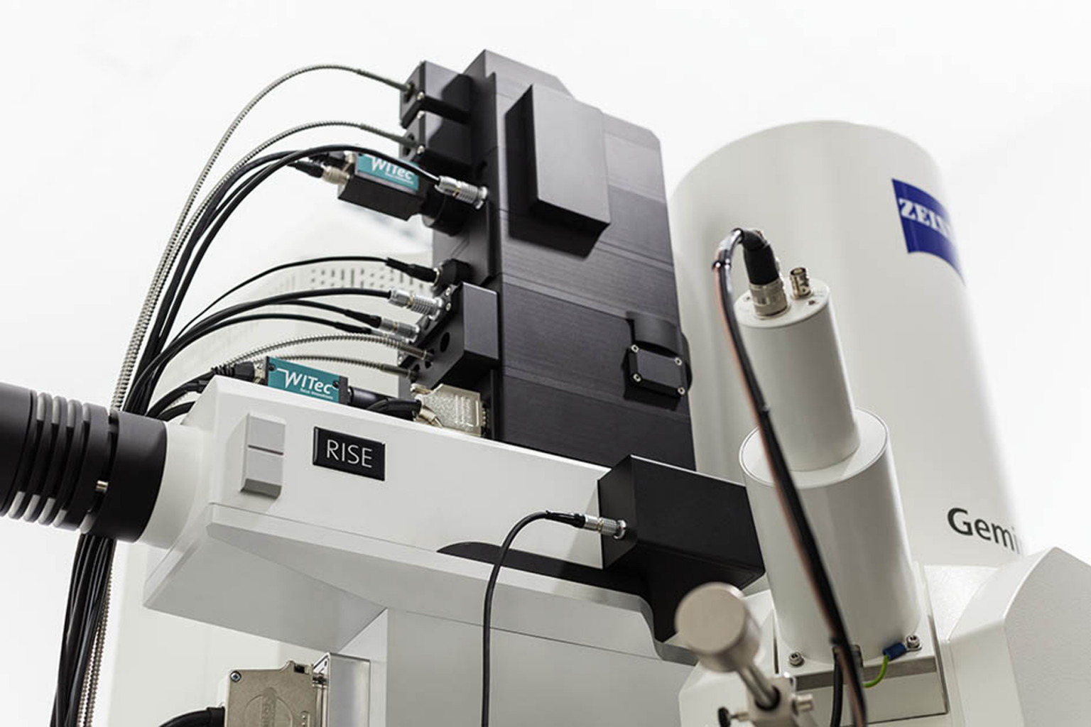
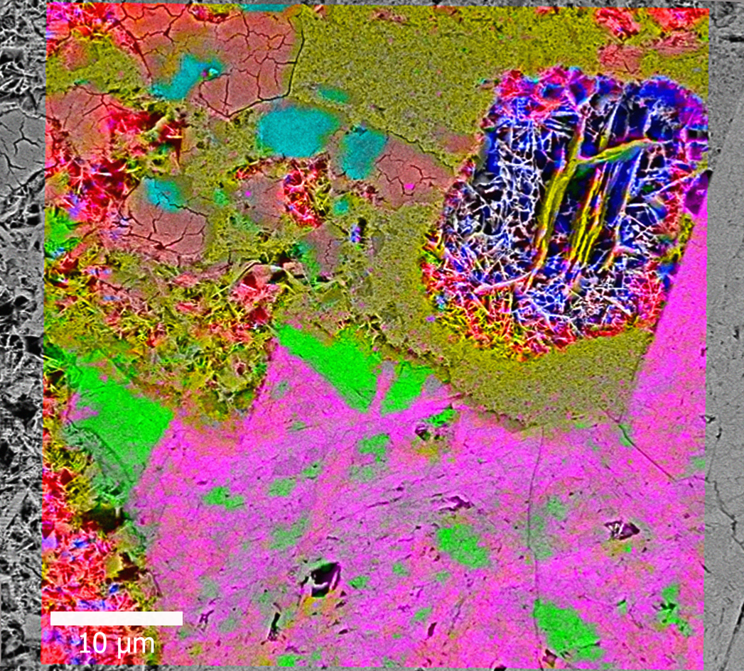
Correlative Raman Sem Imaging Scientist Live Damon strom explores comprehensive particle analysis with correlative raman microscopy. raman microscopy provides exceptional chemical sensitivity that allows data acquisition from very small material volumes, such as those encountered in microparticle analysis. The newly developed correlative rise (raman imaging and scanning electron) microscope enables for the first time the acquisition of sem and confocal raman images within a single instrument (figure 1). this article gives an overview of this new correlative imaging technique and provides two example applications. materials and methods. In this minireview, we summarize the principle of raman and the development of a correlative sem raman system and showcase the typical application of such rise microscopy in different fields. on this basis, the advantages of the rise system, comprising two complementary techniques, are highlighted. Confocal raman imaging and scanning electron (rise) microscopy, when combined in a microscope, complement each other and pro vide the emerging opportunities to clarify morphological, struc tural, and chemical information of materials at the micron and even nanoscale.

Compound Semiconductor Analysis With Correlative Raman Imaging In this minireview, we summarize the principle of raman and the development of a correlative sem raman system and showcase the typical application of such rise microscopy in different fields. on this basis, the advantages of the rise system, comprising two complementary techniques, are highlighted. Confocal raman imaging and scanning electron (rise) microscopy, when combined in a microscope, complement each other and pro vide the emerging opportunities to clarify morphological, struc tural, and chemical information of materials at the micron and even nanoscale. Witec rise microscopy mode for correlative raman sem imaging is now compatible with the zeiss merlin sem. the hybrid system provides raman spectroscopic imaging and advanced ultrastructural analysis, for nanotechnology, life sciences, geosciences, pharmaceutics, materials research and more. The basic of confocal raman and sem imaging; how to integrate raman and sem into one instrument; how to differentiate materials with the same elemental composition; about applications from materials and earth sciences requiring correlative, nondestructive analysis techniques for their full characterization this webinar is presented by witec. Correlative raman sem imaging (rise microscopy, witec, ulm, germany and tescan orsay holding, brno, czech republic) combines an sem and a confocal raman microscope. the confocal raman microscope is integrated into the vacuum chamber of the electron microscope. Analysis of an artificial microbial community demonstrated that our correlative approach was able to resolve the activity of single cells using heavy water sip in conjunction with raman and or.
