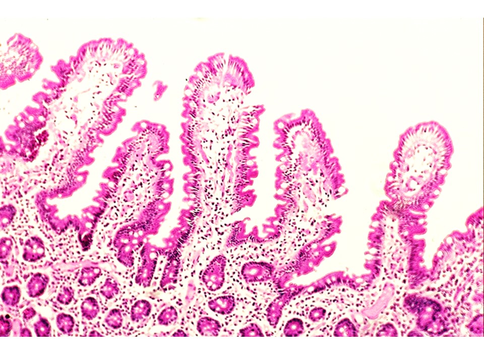
Histology Of Gastrointestinal Tract And Hepatobiliary System Download On upper gi endoscopy, there was duodenal nodularity. biopsy is taken with the clinical suspicion of celiac disease. the histopathological section image is given above. Gastrointestinal, patoloji, atlas, pathology, whole slide image atlas of pathology with whole slide images. 32 duodenum. 33 colon. 35 liver transplantation. 36 benign liver tumors. 37 malignant liver tumors. pancreatobiliary system. 38 pancreas tumors. 39 gallbladder. 40 ampulla of vater. lung. 41 lung tumors.

A Histology Tour Of The Gi Tract The Duodenum Giardiasis is a rare infection, classically found in the duodenum. it can mimic celiac disease. it is also known as beaver fever. clinical features usually two or more of the following: [1] diarrhea x5 days. flatulence. foul smelling feces. nausea. abdominal cramps. excessive tiredness. epidemiology: uncommon. etiology:. The duodenum (about 30 cm long) is the first section of the small intestine that connects the pyloric orifice with the jejunum. it can be divided into superior (d1), descending (d2), inferior (d3) and ascending (d4). This text atlas is one of elsevier’s foundations in diagnostic pathology series, a region specific series of pathology texts intending to provide affordable and up to date presentations of the most essential information required on the diagnostic entities commonly encountered in practice. Book title: atlas of gastrointestinal pathology. book subtitle: as seen on biopsy. authors: i. m. p. dawson. series title: current histopathology. doi: doi.org 10.1007 978 94 009 6583 6. publisher: springer dordrecht. ebook packages: springer book archive. copyright information: i. m. p. dawson 1983.

A Histology Tour Of The Gi Tract The Duodenum This text atlas is one of elsevier’s foundations in diagnostic pathology series, a region specific series of pathology texts intending to provide affordable and up to date presentations of the most essential information required on the diagnostic entities commonly encountered in practice. Book title: atlas of gastrointestinal pathology. book subtitle: as seen on biopsy. authors: i. m. p. dawson. series title: current histopathology. doi: doi.org 10.1007 978 94 009 6583 6. publisher: springer dordrecht. ebook packages: springer book archive. copyright information: i. m. p. dawson 1983. The digestive system can be divided into the digestive tract (oral cavity, esophagus, stomach, small intestine, and large intestine) and associated digestive organs (salivary glands, pancreas, liver, and gallbladder). Aims. giardia: is sometimes missed by the pathologist, and we sought to determine how often this occurs at our institution a large tertiary care center with a subspecialty gastrointestinal pathology service and what certain clinical and histologic clues can be used to flag cases with a higher likelihood of infection, targeting them for greater. Gastrointestinal and liver histology pathology atlas sunday, 10 august 2014. duodenum : giardiasis in this picture, we can oberve the pear shaped giardia trophozoit, in frontal and lateral view: posted by fer at 08:46. email this blogthis! share to x share to facebook share to pinterest. The digestive system consists of the digestive tract—oral cavity, esophagus, stomach, small and large intestines, and anus—and its associated glands—salivary glands, liver, and pancreas (figure 15–1).

A Histology Tour Of The Gi Tract The Duodenum The digestive system can be divided into the digestive tract (oral cavity, esophagus, stomach, small intestine, and large intestine) and associated digestive organs (salivary glands, pancreas, liver, and gallbladder). Aims. giardia: is sometimes missed by the pathologist, and we sought to determine how often this occurs at our institution a large tertiary care center with a subspecialty gastrointestinal pathology service and what certain clinical and histologic clues can be used to flag cases with a higher likelihood of infection, targeting them for greater. Gastrointestinal and liver histology pathology atlas sunday, 10 august 2014. duodenum : giardiasis in this picture, we can oberve the pear shaped giardia trophozoit, in frontal and lateral view: posted by fer at 08:46. email this blogthis! share to x share to facebook share to pinterest. The digestive system consists of the digestive tract—oral cavity, esophagus, stomach, small and large intestines, and anus—and its associated glands—salivary glands, liver, and pancreas (figure 15–1).
