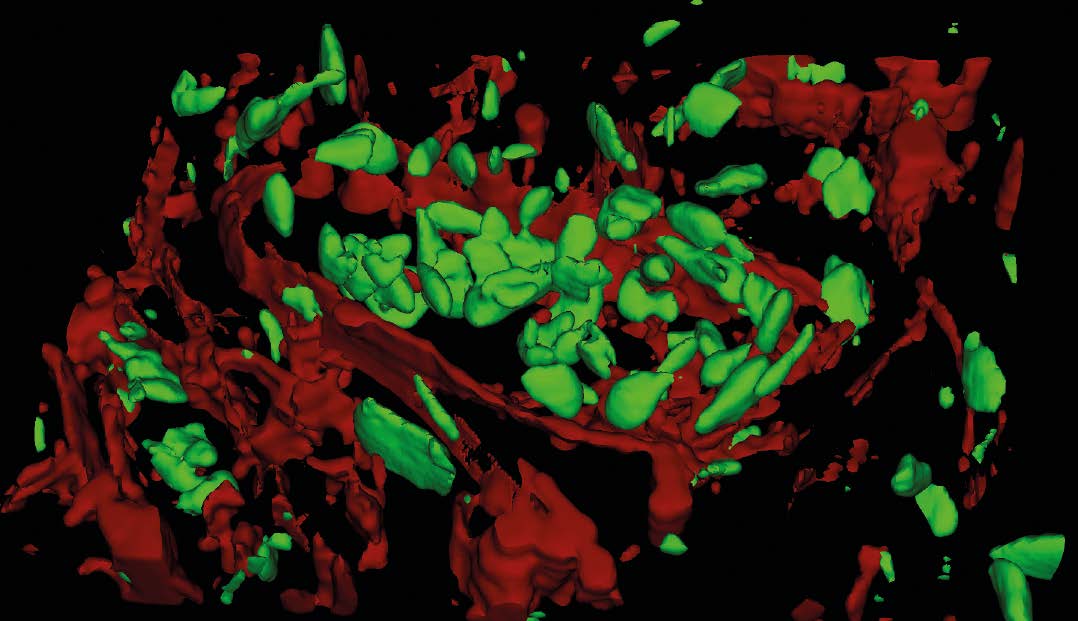
Raman Microscopy For Dynamic Molecular Imaging Of Living Cells Download scientific diagram | microscopy image (a) and raman image (b) constructed based on the cluster analysis (ca) method; raman images of all clusters identified by. Integrating the raman spectrometer with a light microscope ofers a unique ability to noninvasively characterize chemically complex and spatially inhomogeneous samples with a sub micron spatial resolution.

Microscopy Image A And Raman Image B Constructed Based On The Here, we developed a simple raman spectroscopic method that utilized a silver nanoforest (snf) substrate and a hand held raman spectrometer for identifying cancer protein biomarkers with very. Coherent raman scattering (crs) microscopy is gaining acceptance as a valuable addition to the imaging toolset of biological researchers. optimal use of this label free imaging technique benefits from a basic understanding of the physical principles and technical merits of the crs microscope. This review article will provide a brief summary of raman spectroscopy imaging, which includes the use of coherent anti stokes raman spectroscopy (cars), surface enhanced raman spectroscopy (sers), and single walled carbon nanotubes (swnts). Schematic of typical raman hyperspectral imaging systems. (a) ccd based raman microscope for microscopic imaging. (b) line scan raman imaging system for acquiring images from bulk samples. point scanning illuminate the sample with a single spot to acquire a single raman spectrum.

Raman Microscopy This review article will provide a brief summary of raman spectroscopy imaging, which includes the use of coherent anti stokes raman spectroscopy (cars), surface enhanced raman spectroscopy (sers), and single walled carbon nanotubes (swnts). Schematic of typical raman hyperspectral imaging systems. (a) ccd based raman microscope for microscopic imaging. (b) line scan raman imaging system for acquiring images from bulk samples. point scanning illuminate the sample with a single spot to acquire a single raman spectrum. We have demonstrated chemical imaging from the fingerprint and ch stretch regions of the raman spectrum using b cars microscopy and shown at least a fivefold improvement in acquisition speed (50 ms pixel) over spontaneous confocal raman microspectroscopy. Figure 3 shows the optical image (a), raman images (b, c) and raman spectra (d) obtained from a die on si wafer after thermal processing of ni thin films deposited on the substrate. the optical and spectral acquisitions were made using a 100x metallurgical objective having a 0.9 numerical aperture. the raman image in figure 3b was generated as. Comparison of a bright field microscopy image of a mouse brain with stimulated raman scattering (srs) microscopy; dashed line indicates the tumor margin. (b d) high magnification srs images within the tumor (b), at the tumor brain interface (c) and within normal brain (d). In this report, the essential components and features of a modern confocal raman microscope are reviewed using the instance of thermo scientific dxrxi raman imaging microscope, and examples of the potential applications of raman microscopy and imaging for constituents of biosensors are presented.
