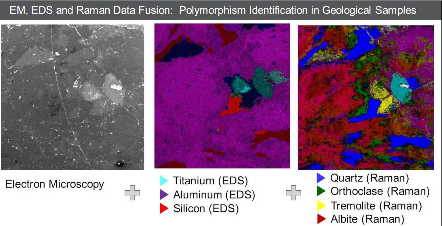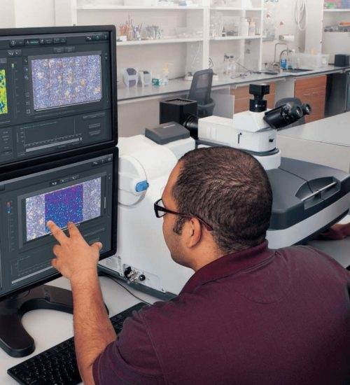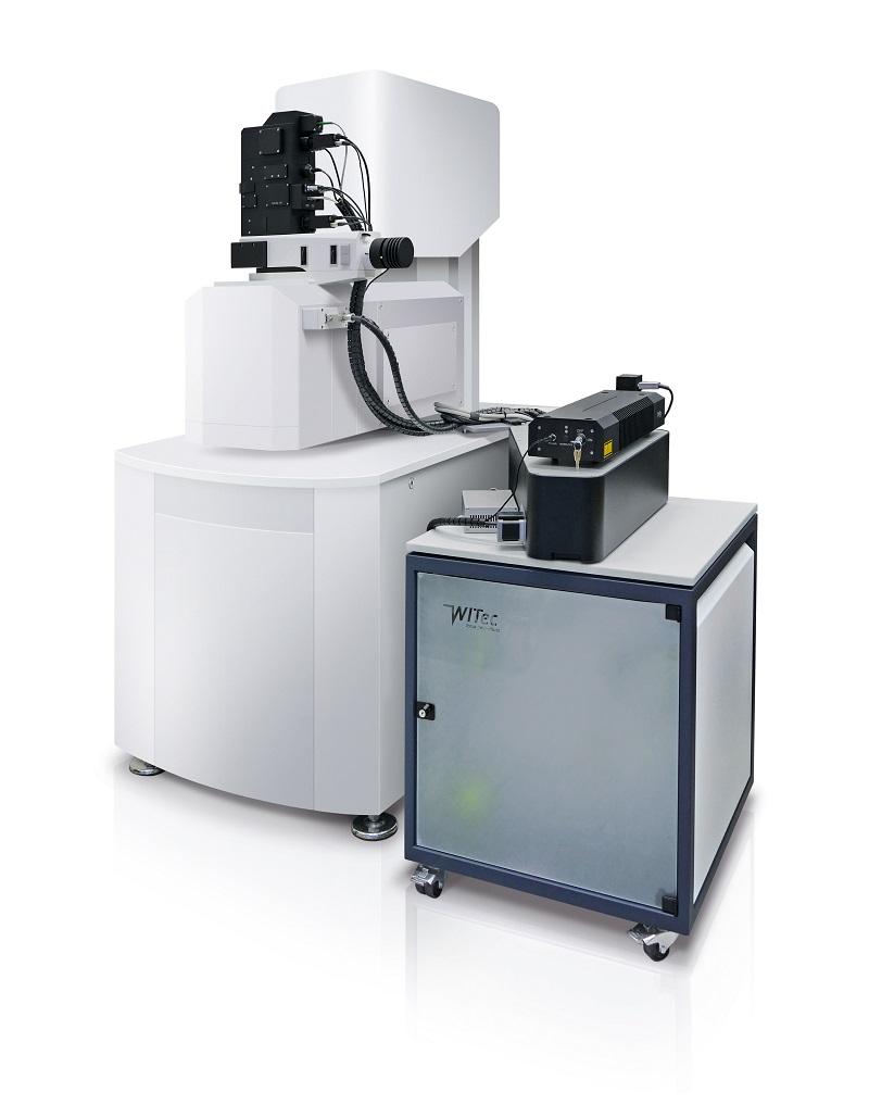
Raman Sem Combined Microscopy Nicoletcz Combination of eds electron microscopy and raman spectroscopy. add raman imaging microscopy to take your results to the next level: samples can be analyzed at once with sem eds and raman microscopy to reveal molecular details of 2d and 3d structures. The user proven dxr raman microscope is now available in the new version of the dxr3xi with a high performance emccd detector and a microscopic table with the possibility of nanoslift for super fast chemical imaging of your samples.

Raman Sem Combined Microscopy Nicoletcz The technique combines sem and raman microscopy, a mutually beneficial pairing, allowing not only the high resolution morphological information obtained by sem but also the chemical and structural analyses of specified areas by raman. Raman imaging and scanning electron microscopy (rise) combine the advantage of scanning electron microscope and raman spectroscopy, which can collect the morphology, composition, and structure information in the same micro region of the geological sample in situ. Rise microscopy is a novel correlative microscopy technique that combines confocal raman imaging and scanning electron (rise) microscopy within one microscope system (fig. 1a). this unique combinat. Samples can be analyzed in parallel with sem eds by raman microscopy to reveal molecular details from 2d and 3d structures. cover the chemical image of your sem structures up to coencoded tag open1 microns with single point measurement or < fast imaging of large areas with the patented thermo scientific maps correlative microscopy software.

08 Simultaneous Two Color Stimulated Raman Scattering Micros Pdf Rise microscopy is a novel correlative microscopy technique that combines confocal raman imaging and scanning electron (rise) microscopy within one microscope system (fig. 1a). this unique combinat. Samples can be analyzed in parallel with sem eds by raman microscopy to reveal molecular details from 2d and 3d structures. cover the chemical image of your sem structures up to coencoded tag open1 microns with single point measurement or < fast imaging of large areas with the patented thermo scientific maps correlative microscopy software. Due to the specific vacuum requirements for scanning electron microscopy (sem), the raman microscope has to operate in vacuum in a correlative raman sem, which is a type of microscope combination that has recently increased in popularity. this works considers the implications of conducting raman mic …. Proč kombinovat ramanovu a elektronovou mikroskopii (sem)? elektronovou mikroskopií získáte obrázky vzorků s velmi vysokým prostorovým rozlišení a využitím tzv. eds detektoru i detailní prvkovou (elementární) mapu vašeho vzorku. spojení elektronové mikroskopie eds a ramanovy spektroskopie. Sem imaging (combined with microanalytical techniques such as eds, wds or ebsd) and micro raman spectroscopy have been combined for materials characterization in several recent studies. switching from one to the other is often considered to be problematic. Correlative raman sem imaging (rise microscopy, witec, ulm, germany and tescan orsay holding, brno, czech republic) combines an sem and a confocal raman microscope. the confocal raman microscope is integrated into the vacuum chamber of the electron microscope. non destructive raman and sem measurements are consecutively performed at.

Correlative Raman Imaging And Scanning Electron Microscopy Raman Sem Due to the specific vacuum requirements for scanning electron microscopy (sem), the raman microscope has to operate in vacuum in a correlative raman sem, which is a type of microscope combination that has recently increased in popularity. this works considers the implications of conducting raman mic …. Proč kombinovat ramanovu a elektronovou mikroskopii (sem)? elektronovou mikroskopií získáte obrázky vzorků s velmi vysokým prostorovým rozlišení a využitím tzv. eds detektoru i detailní prvkovou (elementární) mapu vašeho vzorku. spojení elektronové mikroskopie eds a ramanovy spektroskopie. Sem imaging (combined with microanalytical techniques such as eds, wds or ebsd) and micro raman spectroscopy have been combined for materials characterization in several recent studies. switching from one to the other is often considered to be problematic. Correlative raman sem imaging (rise microscopy, witec, ulm, germany and tescan orsay holding, brno, czech republic) combines an sem and a confocal raman microscope. the confocal raman microscope is integrated into the vacuum chamber of the electron microscope. non destructive raman and sem measurements are consecutively performed at.
