The Impact Of Spatial Normalization For Functional Magnetic Resonance

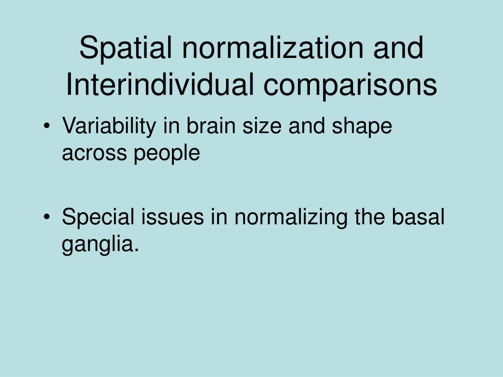
Pdf The Impact Of Spatial Normalization For Functional Magnetic Spatial normalization—the process of aligning anatomical or functional data acquired from different individuals to a common stereotaxic atlas—is routinely used in the vast majority of functional neuroimaging studies, with important consequences for scientific inference and reproducibility. Previous resting state functional magnetic resonance imaging (rs fmri) studies frequently applied the spatial normalization on fmri time series before the calculation of temporal features (here referred to as “prenorm”).

Pdf Landmark Guided Region Based Spatial Normalization For Functional We presented a novel region‐based volumetric spatial normalization solution for human structural and functional brain image processing and statistical analysis. Our results indicated that prenorm may induce algorithmic intersubject variability on tsnr and reduce its reliability, which also significantly affected alff and reho. we suggest using postnorm. During spatial normalization the topology of a brain's anatomical structures should be preserved between healthy normal subjects, with connected neighboring morphological structures remain connected during deformation. The alignment accuracy and impact on functional maps of four spatial normalization procedures have been compared using a set of high resolution brain mris and functional pet volumes acquired in 20 subjects.

Ppt Functional Magnetic Resonance Imaging Powerpoint Presentation During spatial normalization the topology of a brain's anatomical structures should be preserved between healthy normal subjects, with connected neighboring morphological structures remain connected during deformation. The alignment accuracy and impact on functional maps of four spatial normalization procedures have been compared using a set of high resolution brain mris and functional pet volumes acquired in 20 subjects. Spatial normalization—the process of aligning anatomical or functional data acquired from different individuals to a common stereotaxic atlas—is routinely used in the vast majority of. Previous resting state functional magnetic resonance imaging (rs fmri) studies frequently applied the spatial normalization on fmri time series before the calculation of temporal features (here referred to as “prenorm”). Here, we present the first methods for spatial normalization and nonrigid motion correction of fmri data spanning the entire brainstem and cervical spinal cord. Spatial normalization of brains to a standardized space is a widely used approach for group studies in functional magnetic resonance imaging (fmri) data. commonly used template based approaches are complicated by signal dropout and distortions in echo planar imaging (epi) data.

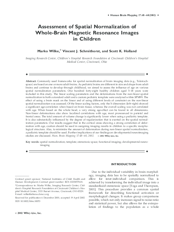
Pdf Assessment Of Spatial Normalization Of Whole Brain Magnetic Spatial normalization—the process of aligning anatomical or functional data acquired from different individuals to a common stereotaxic atlas—is routinely used in the vast majority of. Previous resting state functional magnetic resonance imaging (rs fmri) studies frequently applied the spatial normalization on fmri time series before the calculation of temporal features (here referred to as “prenorm”). Here, we present the first methods for spatial normalization and nonrigid motion correction of fmri data spanning the entire brainstem and cervical spinal cord. Spatial normalization of brains to a standardized space is a widely used approach for group studies in functional magnetic resonance imaging (fmri) data. commonly used template based approaches are complicated by signal dropout and distortions in echo planar imaging (epi) data.

Ppt Functional Magnetic Resonance Imaging Powerpoint Presentation Here, we present the first methods for spatial normalization and nonrigid motion correction of fmri data spanning the entire brainstem and cervical spinal cord. Spatial normalization of brains to a standardized space is a widely used approach for group studies in functional magnetic resonance imaging (fmri) data. commonly used template based approaches are complicated by signal dropout and distortions in echo planar imaging (epi) data.

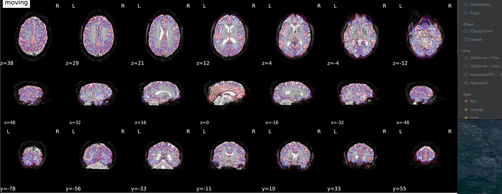
Fmriprep Spatial Normalization Of Functional Images Gone Wrong
Comments are closed.