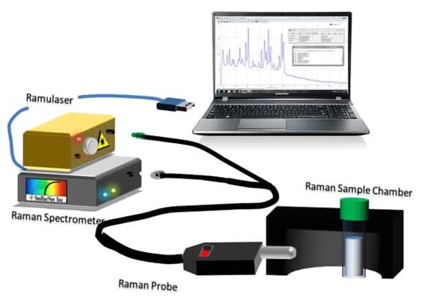
Raman Setup Diagram Stellarnet Inc Each week, we’ll explain the fundamentals of raman spectroscopy step by step in a fun, clear and easy way. check out the previous videos: • raman for beginners | the spectroscop more about. Raman imaging is a technique that generates images with both spectral and spatial information. raman spectra are collected from various spatial positions, and then each spectrum is reduced to only one value for the corresponding pixel. the most common method is using the peak intensity, presenting chemical distribution and concentration.

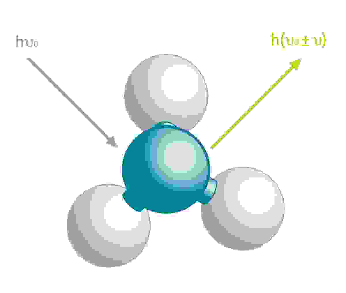
Basic Principles Of Raman Spectroscopy And Its Applications For What is raman imaging? raman imaging, also called raman mapping, is a hyperspectral technique where variations in the raman spectrum across or throughout a sample create a contrast, figure 1. in a raman image, each point contains a full raman spectrum, which is a source of a vast amount of chemical information. In combination with mapping (or imaging) raman systems, it is possible to generate images based on the sample’s raman spectrum. these images show distribution of individual chemical components, polymorphs and phases, and variation in crystallinity. raman spectroscopy is both qualitative and quantitative. Raman microscopy allow samples to be examined visually with the microscope, and then analyzed with raman spectroscopy with the laser. from there, detailed chemical images can even be created. raman microcopy and imaging is an important set of techniques at bruker, so be sure to check out those dedicated websites to learn more. What can raman spectroscopy tell you? learn how raman spectroscopy can reveal the chemistry and structure of materials. we describe the features of a raman spectrum and explain the variations in raman band parameters. we also provide examples of how raman spectra can tell apart different chemicals and polymorphs. raman spectra.

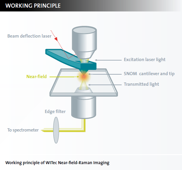
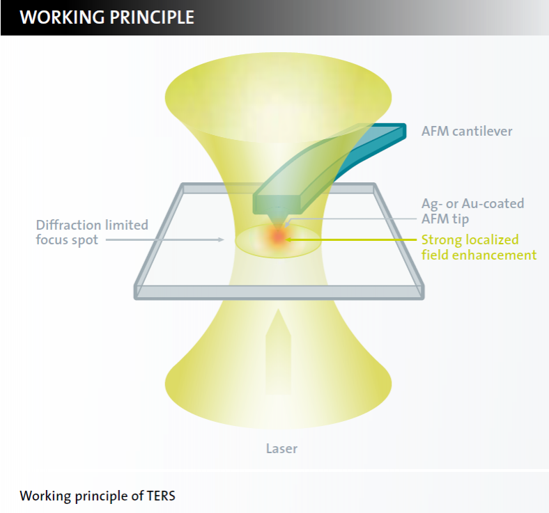
Raman Imaging Witec Raman Imaging Oxford Instruments Raman microscopy allow samples to be examined visually with the microscope, and then analyzed with raman spectroscopy with the laser. from there, detailed chemical images can even be created. raman microcopy and imaging is an important set of techniques at bruker, so be sure to check out those dedicated websites to learn more. What can raman spectroscopy tell you? learn how raman spectroscopy can reveal the chemistry and structure of materials. we describe the features of a raman spectrum and explain the variations in raman band parameters. we also provide examples of how raman spectra can tell apart different chemicals and polymorphs. raman spectra. Where is raman microscopy best applied? how long does it take to measure a raman spectrum? this time we explain some more practical aspects of raman spectros. This tutorial will teach introductory raman spectroscopy and imaging. the chemical bond origins of raman scattering along with the instrumentation used to acquire raman spectra and images will be explained. in particular, we will discuss the selection of laser excitation wavelength, lateral and axial spatial resolution, detection limits, and. In raman microscopy, a research grade optical microscope is coupled to the excitation laser and the spectrometer. this produces images down to 1 micron and generates raman spectra. imaging and spectroscopy can be combined to generate raman cubes which are three dimensional data sets, yielding spectral information at every pixel of the 2d image. Can you perform particle characterization using raman microscopy? how to know if a raman system is confocal? can you perform raman measurements in controlled environment? what are the best modes for fast raman mapping? what are the main and unique features of labram™ soleil? how do you obtain the best raman spectral resolution?.

Raman Imaging Witec Raman Imaging Oxford Instruments Where is raman microscopy best applied? how long does it take to measure a raman spectrum? this time we explain some more practical aspects of raman spectros. This tutorial will teach introductory raman spectroscopy and imaging. the chemical bond origins of raman scattering along with the instrumentation used to acquire raman spectra and images will be explained. in particular, we will discuss the selection of laser excitation wavelength, lateral and axial spatial resolution, detection limits, and. In raman microscopy, a research grade optical microscope is coupled to the excitation laser and the spectrometer. this produces images down to 1 micron and generates raman spectra. imaging and spectroscopy can be combined to generate raman cubes which are three dimensional data sets, yielding spectral information at every pixel of the 2d image. Can you perform particle characterization using raman microscopy? how to know if a raman system is confocal? can you perform raman measurements in controlled environment? what are the best modes for fast raman mapping? what are the main and unique features of labram™ soleil? how do you obtain the best raman spectral resolution?.

Raman Imaging Witec Raman Imaging Oxford Instruments In raman microscopy, a research grade optical microscope is coupled to the excitation laser and the spectrometer. this produces images down to 1 micron and generates raman spectra. imaging and spectroscopy can be combined to generate raman cubes which are three dimensional data sets, yielding spectral information at every pixel of the 2d image. Can you perform particle characterization using raman microscopy? how to know if a raman system is confocal? can you perform raman measurements in controlled environment? what are the best modes for fast raman mapping? what are the main and unique features of labram™ soleil? how do you obtain the best raman spectral resolution?.
